
Mini Review / Open Access
DOI: 10.31488/jjm.1000107
Diagnosis of Cancer Using X-ray Diffraction of Skin
Veronica J. James
Research School of Chemistry, Australian National University, Canberra, ACT, Australia
*Corresponding author:Veronica J. James, Research School of Chemistry, Australian National University, Canberra, ACT, Australia
Introduction
Skin samples can be used to diagnose a number of cancers (such as melanoma, high and low grade prostate cancer, bowel) by simply spotting the different additions to the skin diffraction pattern made by each cancer. Diffraction patterns of any tissue simply record every atom in the tissue, a physical fact. Hence as each cancer produces a unique change to the skin pattern of a patient, as soon as it starts, the only conclusion is that a new molecule has been added to the skin for all patients with that particular cancer type. All that is required is the identification of any changes that appear in the particular patient’s skin pattern. The diagnosis of each sample becomes as simple as a child’s game of “Spot The Difference”.
In order to understand how such diagnostic tests work, we need firstly to observe the normal diffraction pattern for skin [1-3], shown in figure 1.
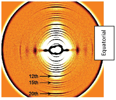
Figure 1.Normal skin diffraction pattern. Colours in the pattern are computer enhanced for easier viewing
Three of the strong meridional reflections have been numbered as all changes occur in the region between these reflections. The lower meridional reflections may or may not be present depending on the machine setup. For skin research, all that is needed is a 3mm skin biopsy which can be taken from anywhere on the body using a Keyes punch. Our samples are usually taken from the softer skin on the stomach or buttocks. This biopsy is immediately immersed in saline and kept frozen at -20˚.
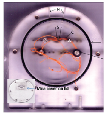
Figure 2.Cell used for skin studies. Sample (S) is held in place by two strands of cotton. Holes(H) allow water to be inserted into the cell, after it is sealed by the O ring, to maintain humidity, and T are the screws for maintaining tension in the sample.
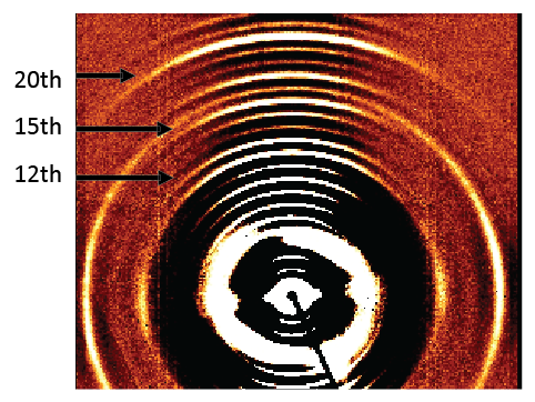
Figure 3.Diffraction pattern with melanoma change
If the samples are taken from melanoma patients, the subsequent diffraction pattern for these samples is found to be altered from that in figure 1. The altered skin diffraction pattern now has full ring that passes through the 16th meridional order but which is stronger in the equatorial direction. This ring has been identified as being associated with every melanoma sample [4]. This melanoma skin diffraction pattern is shown in Figure 3, where the added ring is indicated by a blue arrow. The ring pattern that was added by the melanoma stands out, it is the only change. The presence of this extra ring in the diffraction pattern can be used as a standard test for the presence of melanoma in any patient. All that is needed is a 3mm skin biopsy.
A full diffraction study of high or low grade prostate cancer covering 410 samples supplied by urologists in Australia, Europe, and the USA has shown that prostate cancers cause different changes in the diffraction patterns at every stage. These results indicated that the prostatectomies in 70% of the patients occurred too late as the cancer had already invaded into the body either before or during the procedures, whether this was a robotic procedure or not.
The diffraction patterns obtained for all biopsy skin samples collected from low grade prostate cancer patients is shown in figure 7. The arrow indicates an additional circle, which lies between the 13th and 14th meridional orders. This ring is the change from the normal skin pattern, if the patient is only suffering from a low grade prostate cancer. This change is added to the diffraction pattern of skin by the prostate cancer itself as soon as the cancer starts. This fact has been verified using Tramp mice [5].
Some patients are concerned when the prostate is enlarged and the doctor diagnoses BPH, or benign prostatic hyperplasia or hypertrophy. The skin diffraction test results correctly diagnoses BPH, as it produces a different change in the skin pattern from that of low grade prostate cancer. The skin biopsy of a prostatic cancer patient, who has BPH, would show a ring of change with a larger diameter than that of a prostate cancer patient. For a patient with only BPH, the ring of change lies between the fifteen and sixteen orders as shown in figure 8.
However, should both of these conditions occur for the same patient, both these rings will be added to the resulting diffraction pattern as illustrated in Figure 9. A low grade prostate cancer, if not successfully treated, will change into a high grade prostate cancer. The ring of change gradually increases in diameter passing through the 15th meridional at Gleason 7. Unfortunately, by this time, the cancer may have invaded the body Chances of recovery are greatly reduced.
Regrettably all such samples, supplied for my research, came from post prostatectomies and the results of the diffraction study indicated that 70% had already invaded. The diffraction patterns of all high grade cancers show at least the sharp ring denoting perineural invasion on the17th meridional order or both this ring and another sharp ring on the 18th meridional order that denotes lymphatic invasion. Such a high grade prostate cancer pattern is shown in figure 10.
The ring of change now reaches from between the 13th and 14th orders to beyond the 15th order as indicated by the red arrow. The 17th order, denoted by a green arrow, showing that the cancer had already invaded. The diffraction patterns of all high grade cancers show at least the sharp ring denoting a perineural invasion, {PNI}, which is indicated by the ring on the 17th order, marked by green arrow. Invasion of the body via the lymph system {LVI} also occurs and is indicated by the ring on the 18th order. Unfortunately, these invasions are not removed by a radical prostatectomy, they are still clearly visible in figure 8 after the prostate is removed.
The big advantage of the diffraction test is that the change in the diffraction pattern is caused by the cancer as soon as it starts and prostate cancers have been detected in men from Caucasian heritage in their early 20’s and from 18 years in men of Negroid heritage. If these low grade cancers were immediately treated by chemotherapy, the cancer could be cured. A further skin diffraction test would verify the cure, as the change in the skin diffraction pattern will disappear when the cancer is gone. Unfortunately, although two of my prostate cancer patients were shown to have been cured, there were no earlier tests to verify that the cancers had ever existed. So in order to demonstrate this truly remarkable feature of the diffraction tests I am introducing a patient who had a hair diffraction test.
This lady had a lumpectomy to remove a small breast cancer. Her doctor then suggested chemotherapy to guarantee that the cancer had been completely removed. This caused her hair to fall out and she sent it all to me. Working back along a hair and knowing the date of the lumpectomy, it was easy to obtain the section of hair 2 days before the lumpectomy. There was no need for this woman to have suffered the chemotherapy and loss of her hair [Figure 9a-c].
Now that X-ray machines are available to carry out these tests in Radiology facilities anywhere, such tests could be the order of the day. Many lives would be saved, many treatments avoided. The skin tests can be done in 60 seconds on these laboratory machines.
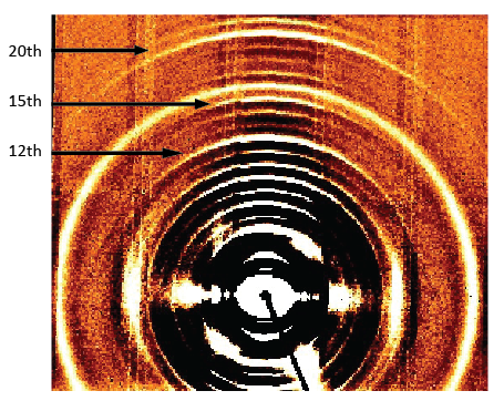
Figure 4.Diffraction pattern with melanoma change
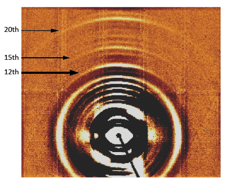
Figure 4B.Diffraction pattern with melanoma change
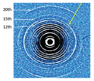
Figure 6.Sample showing the presence of low grade prostate cancer and melanoma
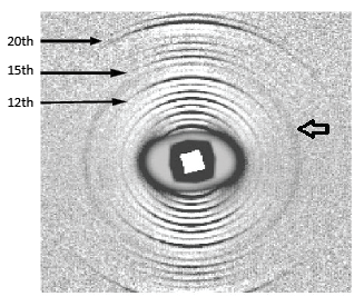
Figure 7.A typical diffraction pattern from a patient who has low grade prostate cancer. The ring of change lies between the 13th and 14th meridional orders and is indicated by an open arrow
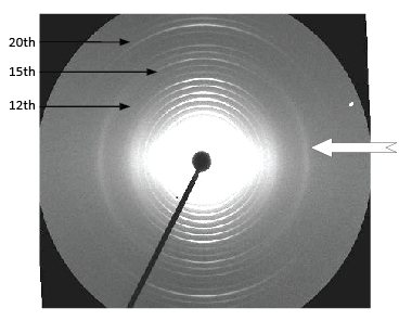
Figure 8.A typical diffraction pattern from a patient who has BPH. The change from a normal skin diffraction pattern is the ring indicated by the arrow
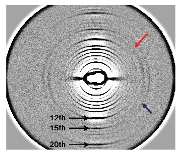
Figure 9.The skin diffraction pattern for a patient with low grade prostate cancer, indicated by a red arrow, and BPH, indicated by a dark blue arrow
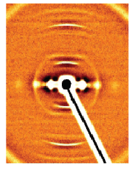
Figure 9a.Illustrates the clear breast cancer diffraction pattern at that time
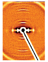
Figure 9b.The pattern 8 days after the lumpectomy, this pattern shows a very much weaker ring of change
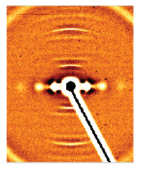
Figure 9c.the pattern 18 days after the lumpectomy, 2 days before the commencement of the chemotherapy, showing no cancer ring thus verifying that the cancer had been completely removed by the lumpectomy
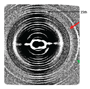
Figure 10.Diffraction pattern from a patient with a high grade prostate cancer, Gleason Score > 7.
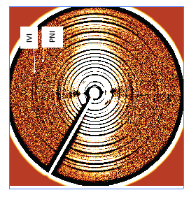
Figure 11.Skin diffraction pattern after radical prostatectomy, both invasions, PNI [perineural] and LVI[via lymph vascular system] are shown to have occurred.
References
Brodsky B, Eikenberry EF Cassidy K. An unusual collagen periodicity in skin. Biochim Biophys Acta Proteins. 1980; 621:162–6.
Stinson RH, Sweeney PR. Skin collagen has an unusual d-spacing. Biochim Biophys Acta Proteins 1980; 621:158–61.
James VJ, Willis BE. Molecular changes in skin predict predisposition to breast cancer JMedGenet. 2002; 39 (2): 1e.
James VJ, Kirby N. The connection between the presence of melanoma and changes in fibre diffraction patterns. Cancers. 2010; 2: 1155-1165.
James VJ, O’Malley Ford JM. Fibre diffraction analysis of skin offers a very early and extremely accurate diagnostic test for prostate cancer. J Cancer Res. 2014; 2014:6
James VJ, O’Malley Ford JM, Buttigieg J. Then there were none. Integr Cancer Sci Therap. 2015; 2(6): 305-307.
Received: June 13, 2018.
Accepted: June 27, 2018.
Published: June 30, 2018.
To cite this article :